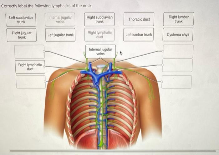
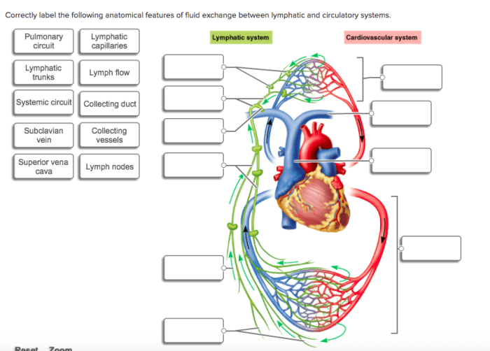
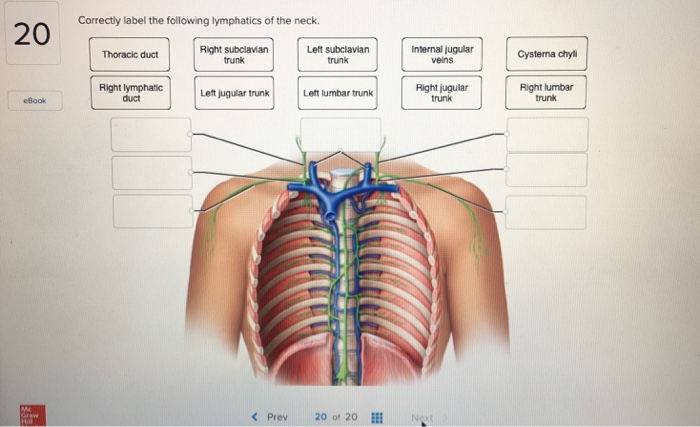
Correctly label the following lymphatics of the thoracic cavity. – Embark on a comprehensive exploration of the lymphatics of the thoracic cavity, unraveling their intricate network and clinical significance. This guide delves into the anatomy, drainage patterns, and clinical implications of these crucial structures, providing a thorough understanding for medical professionals and students alike.
From the thoracic duct, the body’s primary lymphatic vessel, to the intricate mediastinal and tracheobronchial lymph nodes, each lymphatic structure plays a vital role in maintaining fluid balance, immune surveillance, and overall health. This guide empowers you with the knowledge to accurately label and comprehend the functions of these essential components of the lymphatic system.
Lymphatics of the Thoracic Cavity: Correctly Label The Following Lymphatics Of The Thoracic Cavity.

The lymphatics of the thoracic cavity play a crucial role in maintaining fluid balance and transporting immune cells throughout the body. The major lymphatic vessels of this region include the thoracic duct, right lymphatic duct, bronchomediastinal lymph trunks, mediastinal lymph nodes, tracheobronchial lymph nodes, and esophageal lymph nodes.
Thoracic Duct
The thoracic duct is the largest lymphatic vessel in the body. It begins at the cisterna chyli in the abdomen and ascends through the posterior mediastinum to terminate at the left subclavian vein. Along its course, it receives lymph from the lower limbs, pelvis, abdomen, left upper limb, and left side of the head and neck.
- Location: Posterior mediastinum, ascending from the cisterna chyli to the left subclavian vein.
- Tributaries: Lymph from the lower limbs, pelvis, abdomen, left upper limb, and left side of the head and neck.
- Clinical significance: Obstruction of the thoracic duct can lead to chylous ascites, a condition characterized by the accumulation of lymphatic fluid in the peritoneal cavity.
Right Lymphatic Duct
The right lymphatic duct is a smaller lymphatic vessel that drains lymph from the right upper limb and right side of the head and neck. It terminates at the right subclavian vein.
- Location: Right side of the mediastinum, ascending to the right subclavian vein.
- Tributaries: Lymph from the right upper limb and right side of the head and neck.
- Clinical significance: Obstruction of the right lymphatic duct can lead to lymphedema of the right upper limb and right side of the head and neck.
Bronchomediastinal Lymph Trunks, Correctly label the following lymphatics of the thoracic cavity.
The bronchomediastinal lymph trunks are a group of lymphatic vessels that drain lymph from the lungs, bronchi, and mediastinum. They terminate at the thoracic duct.
- Location: Posterior mediastinum, draining into the thoracic duct.
- Tributaries: Lymph from the lungs, bronchi, and mediastinum.
- Clinical significance: Enlargement of the bronchomediastinal lymph trunks can indicate the presence of lung cancer or other mediastinal pathology.
Mediastinal Lymph Nodes
The mediastinal lymph nodes are a group of lymph nodes located in the mediastinum. They receive lymph from the lungs, bronchi, esophagus, and heart.
- Location: Mediastinum, organized into anterior, middle, and posterior groups.
- Drainage patterns: Lymph from the lungs, bronchi, esophagus, and heart.
- Clinical significance: Enlargement of the mediastinal lymph nodes can indicate the presence of infection, cancer, or other mediastinal pathology.
Tracheobronchial Lymph Nodes
The tracheobronchial lymph nodes are a group of lymph nodes located around the trachea and bronchi. They receive lymph from the trachea, bronchi, and lungs.
- Location: Around the trachea and bronchi.
- Tributaries: Lymph from the trachea, bronchi, and lungs.
- Clinical significance: Enlargement of the tracheobronchial lymph nodes can indicate the presence of lung cancer or other respiratory pathology.
Esophageal Lymph Nodes
The esophageal lymph nodes are a group of lymph nodes located along the esophagus. They receive lymph from the esophagus and surrounding structures.
- Location: Along the esophagus.
- Drainage patterns: Lymph from the esophagus and surrounding structures.
- Clinical significance: Enlargement of the esophageal lymph nodes can indicate the presence of esophageal cancer or other esophageal pathology.
FAQ Summary
What is the clinical significance of the thoracic duct?
The thoracic duct is a crucial conduit for lymphatic fluid drainage from the body’s lower extremities, abdomen, and left upper limb. Its obstruction can lead to chylothorax, a condition characterized by the accumulation of lymphatic fluid in the pleural space, potentially compromising respiratory function.
How do the mediastinal lymph nodes contribute to immune surveillance?
The mediastinal lymph nodes are strategically located to filter lymphatic fluid draining from the lungs, heart, and mediastinum. They play a vital role in trapping and eliminating pathogens, contributing to the body’s immune defense mechanisms.
What is the drainage pattern of the tracheobronchial lymph nodes?
The tracheobronchial lymph nodes receive lymphatic drainage from the trachea, bronchi, and lungs. They are responsible for filtering and monitoring inhaled particles and pathogens, contributing to the maintenance of respiratory health.


