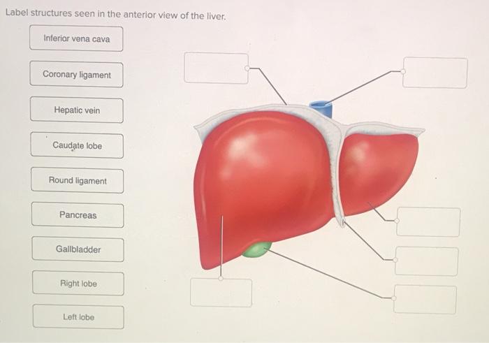
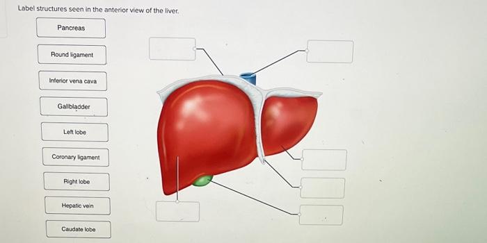
Label structures seen in the anterior view of the liver provide a comprehensive understanding of the liver’s anatomy, essential for surgical and medical procedures. This guide offers a detailed exploration of the liver’s anatomical landmarks, fissures, lobes, segments, vascular structures, and biliary system, empowering readers with a thorough knowledge of this vital organ.
The liver, located in the upper right quadrant of the abdominal cavity, is the largest internal organ, playing a crucial role in metabolism, detoxification, and bile production. Its anterior surface presents distinct anatomical features that serve as important reference points for surgeons and clinicians.
Anatomical Landmarks of the Anterior Liver

The liver is located in the right upper quadrant of the abdominal cavity, beneath the diaphragm. It is the largest internal organ in the human body, weighing approximately 1.5 kilograms. The liver has a wedge-shaped appearance, with its base directed upward and to the right, and its apex pointing downward and to the left.
The anterior surface of the liver is smooth and convex, and it is covered by the peritoneum. The posterior surface of the liver is irregular and concave, and it is attached to the diaphragm, the stomach, and the duodenum by ligaments.
Adjacent Organs and Structures
- Diaphragm
- Stomach
- Duodenum
- Gallbladder
- Right kidney
- Adrenal gland
- Pancreas
Major and Minor Fissures of the Liver: Label Structures Seen In The Anterior View Of The Liver

Major Fissures
The liver is divided into two main lobes, the right and left lobes, by the falciform ligament. The falciform ligament is a thick band of connective tissue that extends from the anterior abdominal wall to the liver. The right lobe is larger than the left lobe and it occupies most of the right upper quadrant of the abdominal cavity.
The left lobe is located to the left of the falciform ligament and it occupies the left upper quadrant of the abdominal cavity.
The liver is also divided into four segments by the portal vein and the hepatic artery. The segments are numbered I to IV, with segment I being located in the right lobe and segments II, III, and IV being located in the left lobe.
Minor Fissures
In addition to the major fissures, there are also a number of minor fissures that divide the liver into smaller segments. These fissures are not as deep as the major fissures and they do not extend all the way through the liver.
The minor fissures include the umbilical fissure, the ligamentum teres fissure, and the porta hepatis fissure.
Liver Lobes and Segments
Lobes
- Right lobe
- Left lobe
- Caudate lobe
- Quadrate lobe
Segments, Label structures seen in the anterior view of the liver
| Segment | Lobe |
|---|---|
| I | Right |
| II | Left |
| III | Left |
| IV | Left |
| V | Right |
| VI | Right |
| VII | Right |
| VIII | Right |
Vascular Structures in the Anterior Liver
Portal Vein
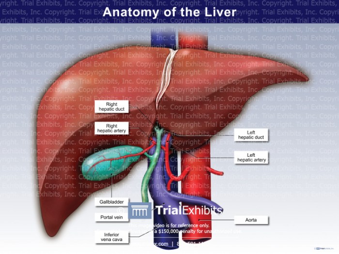
The portal vein is a large vein that carries blood from the intestines, pancreas, and spleen to the liver. The portal vein enters the liver at the porta hepatis and it divides into two branches, the right and left portal veins.
The right and left portal veins supply blood to the right and left lobes of the liver, respectively.
Hepatic Artery
The hepatic artery is a branch of the celiac trunk that supplies oxygenated blood to the liver. The hepatic artery enters the liver at the porta hepatis and it divides into two branches, the right and left hepatic arteries. The right and left hepatic arteries supply blood to the right and left lobes of the liver, respectively.
Hepatic Veins
The hepatic veins are three large veins that carry blood from the liver to the inferior vena cava. The hepatic veins exit the liver at the posterior surface and they join the inferior vena cava just below the diaphragm.
Biliary System in the Anterior Liver

The biliary system is a network of ducts that carry bile from the liver to the duodenum. Bile is a fluid that is produced by the liver and it helps to digest fats. The biliary system consists of the hepatic ducts, the common hepatic duct, and the common bile duct.
The hepatic ducts are small ducts that collect bile from the liver cells. The hepatic ducts join together to form the common hepatic duct. The common hepatic duct then joins with the cystic duct, which carries bile from the gallbladder, to form the common bile duct.
The common bile duct carries bile to the duodenum.
Question Bank
What are the major fissures of the liver?
The major fissures of the liver are the longitudinal fissure and the transverse fissure, which divide the liver into right and left lobes, and superior and inferior segments, respectively.
What is the significance of liver segments?
Liver segments are important for surgical planning, as they allow for precise resection of specific liver areas while preserving healthy tissue.
What is the function of the hepatic portal vein?
The hepatic portal vein carries nutrient-rich blood from the intestines to the liver for processing and detoxification.
